headerimage

We are an ARUP-owned core lab serving the mission of Huntsman Cancer Institute to further cancer research. Our services are available for all University of Utah-associated laboratories at a discounted rate. Outside of the University of Utah, our services are available on an individual basis.

New clients are required to set up an account before submitting specimens to ARUP Research Histology. See below for more information.


Specimens can be placed in our drop-off box during business hours. If you have fresh tissue for a frozen project, please email the lab (aphistologyresearch438@aruplab.com) to coordinate a time to drop off the tissue.

Research Histology does not provide supplies, gross specimens, cassette samples, or transfer formalin-fixed tissue to 70% ethanol in which we require all projects to be submitted.

For pricing information, please contact Heather Homer (heather.homer@aruplab.com).

Research Histology—ARUP Laboratories
Huntsman Cancer Hospital

1950 Circle of Hope Drive, Room N3970
Salt Lake City, UT 84112
Monday–Friday, 8 a.m.–4p.m.

Phone: 801-213-4272
Email: APHistologyResearch438@aruplab.com

Laboratory

For immunohistochemical staining, please contact BMP-ResearchIHC@hci.utah.edu.

 
Client ID Account Setup

All clients requesting services from Research Histology, and/or approval for immunohistochemical staining through ARUP, are required to set up an ARUP Client ID (account). Please follow the detailed instructions below to ensure we are in compliance with the regulatory agencies that govern our operations.

For questions regarding account setup information, contact the ARUP Clinical Trials department (clinicaltrials@aruplab.com). For technical questions, contact Research Histology (APHistologyResearch438@aruplab.com).

ANIMAL SPECIMENS

If you are working with animal samples only, the process for setting up an ARUP Client ID takes approximately 8–10 business days.

Please fill out and submit the following form.

Once your account setup is complete, you will receive a custom ARUP requisition to submit your specimens to Research Histology.

HUMAN SPECIMENS

If your project requires the submission of HUMAN specimens, you must apply for an exemption from electronic (EPIC) ordering. Please follow the instructions below:

To request an ARUP electronic (EPIC) ordering exemption or the creation of an alias medical record, please save the provided Exclusion Research Data Procedure (PDF) form with the IRB number in the file name. Complete the form and email the Clinical Research Compliance and Education (CRCE) officer with Compliance Services at the University of Utah. The compliance office will review and respond within five business days.

This procedure is defined by the CRCE office, not ARUP. We cannot set up an account for processing human specimens without this exemption approval.

Once you have gone through the steps above, you will receive a notification that your exemption has been approved. This information, along with the ARUP Client Account Information form, will be used to set up a research account for you at ARUP. Once your account setup is complete, you will receive a custom ARUP requisition to submit your specimens.

Immunohistochemical Staining

If your project requires additional immunohistochemical staining, please contact BMP-ResearchIHC@hci.utah.edu

Services Provided

(Arranged according to the frequency that the service is provided)

Decalcification: Our lab offers decalcification services. All tissue containing calcium (bone, bone marrow, etc.) must be submerged in a decal solution prior to processing and embedding until tissue is pliable. Specimens containing calcium cannot be cut after paraffin embedding. Undecalcified tissue will exhibit cutting artefact and will most likely stain poorly. “Tissue containing calcium must undergo calcium removal before embedding unless specific studies requiring undecalcified bone are requested.” (Carson, 2009, p. 46).

Processing: This automated protocol involves running tissue through a vacuum infiltration processing machine, which replaces all water present in the tissue with paraffin wax to stabilize the tissue and make it amenable to cutting.

Paraffin embedding: This process involves orienting warm tissue samples into hot paraffin wax molds, which are then cooled until the paraffin block is solid. Tissue is embedded in the smallest mold appropriate for the tissue size. Embedding instructions, provided by the client, guide lab staff on how to orient the tissue samples so that the area of interest is sectioned correctly.

Cell blocks: Sometimes collections are of a concentration of cells rather than a piece of tissue. We accept cell aggregate submissions for processing, embedding, and sectioning. Note: Cell aggregate needs to be wrapped in Histowrap; otherwise, cells may be lost during processing.

Shaves/scrolls: Often used for DNA extraction, a shave/scroll is cut in a specific way such that the “section” turns out as a scroll. Shave/scrolls are 10 μm or thicker and delivered in an Eppendorf tube.

Tissue punches: Punches are used in assembling a tissue microarray or for simply examining a small piece of representative tissue. The specimen is delivered in an Eppendorf tube. Accompanying hematoxylin and eosin (H&E)-stained slides with the area of interest circled must be provided.

TMA (tissue microarray): Many 2 mm representative tissue punches are extracted from different paraffin donor blocks and re-embedded into a single recipient (microarray) block at defined array coordinates. A microarray block may contain up to 60 tissue cores. After sectioning, a microarray slide is amenable to high-throughput analysis of multiple specimens at the same time. (Jawhar, 2009, p. 2018)

Repair of broken slides: Slides with clean breaks (not shattered) can be repaired.

Wax-dipped slides: This process curbs oxidization and is performed when slides will be stored prior to testing.

Cutting

(Arranged topically)

Sectioning: After a specimen is embedded into a paraffin wax block, it can be sectioned on a microtome by a histologist. All sections are initially unstained. We can cut as many sections from a block as necessary, up to depletion of the block. Possible ways to section a block include:

  • Levels: A section is obtained from the surface of the block, and then from another plane deeper in the tissue of the block. Specific depth between levels may be requested.
  • Serial sections: Sequential sections are cut, starting at the shallowest area of the tissue and picking up all sections through the entirety of the tissue.
  • Tissue orientation: When tissue elements need to be oriented a specific way on a slide, embedding instructions must include the required orientation. The area of interest needs to be embedded in the bottom of the tissue mold. This will be sectioned first. The surface of the tissue may be indicated by inking the tissue surface to aid in orientation. The embedding instructions would then include to embed the tissue ink side down.

Cryosectioning: Once a fresh specimen is embedded in optimal cutting temperature (OCT) compound, it can be cut on a cryomicrotome (cryostat). Orientation guidelines are parallel to regular microtome sectioning. All sections are initially unstained. Various stains can be applied.

Special Staining

(Arranged alphabetically)

AFB (Acid-Fast Bacilli–Zeil-Neelsen Method)

  • Acid-fast bacteria (e.g., mycobacterium) exhibit acid fastness, that is, they “stand fast to” or resist acid- and/or ethanol-based decolorization during the staining process.
  • Acid fastness is a physical property of the cell. An acid-fast stain is used to identify acid-fast mycobacteria in tissue sections. (Carson, 2009, p. 226)
  • Acid-fast bacteria of interest are pathogenic to humans.
  • In general, acid-fast stains allow bacteria to be divided into acid-fast and nonacid-fast groups. (Carson, 2009, p. 222)
  • 4–5 μm

Alcian Blue, pH 2.5 Stain

  • Used for demonstration of acid mucopolysaccharides (Carson, 2009, p. 145)
  • May be used in the diagnosis of diseases such as mucopolysaccharidosis
  • 4–5 μm

Alcian Blue, with Hyaluronidase Digestion

  • For differentiation of epithelial and connective tissue mucins (Carson, 2009, p. 147)
  • Bovine testicular hyaluronidase digestion highlights mucosubstances containing hyaluronic acid and chondroitin sulfates A and C (Carson, 2009, p. 148)
  • 4–5 μm

Alcian Blue, with PAS

  • For differentiation between neutral and acidic mucosubstances, this procedure is used in many laboratories today for the detection of intestinal metaplasia. (Carson, 2009, p. 148)
  • The alcian blue technique stains the acidic mucosubstances, whereas the PAS stains the neutral mucosubstances. (Carson, 2009, p. 148)
  • 4–5 μm; sections of kidney should be cut at 2–3 μm.

Aniline Blue

  • When used alone, aniline blue is a general stain used for broad differentiation of tissue histology. Hence, it is primarily used for slides to be scraped of tumor for molecular testing.
  • Aniline blue or its constituents are used to stain collagen, as the fiber stain in Masson’s trichrome (HT-PROC-4441).

Auramine Rhodamine Stain

  • Used to detect mycobacterium tuberculosis or other acid-fast organisms. (Carson, 2009, p. 230) While the AFB stain is more specific for acid-fast organisms, auramine rhodamine is more affordable and sensitive and so is frequently used for initial screening. (Kommareddi, 1984)
  • 4–5 μm

Bielschowsky Method Stain

  • Demonstrates nerve fibers and the presence of neurofibrillary tangles and senile plaques in Alzheimer disease. It is typical for some neurofibrils to result as aging occurs, but in Alzheimer’s disease, tremendous numbers of neurofibrils develop. (Carson, 2009, p. 202).
  • 8 μm

Brown-Hopps Modified Gram Stain

  • Used for demonstrating gram-negative and gram-positive bacteria in tissue. (Carson, 2009, p. 231) The walls of gram-positive bacteria retain the initial crystal violet dye, whereas the walls of gram-negative bacteria will decolorize to accept a counterstain. In this way, the two types are differentiated.
  • In general, gram stains allow bacteria to be classified as gram-positive and gram-negative organisms. (Carson, 2009, p. 222)
  • 4–5 μm

Colloidal Iron

  • Demonstrates carboxylated and sulfated mucopolysaccharides and glycoproteins, which absorb colloidal ferric ions at a low pH. (Carson, 2009, p. 149)
  • 4-5 μm

Congo Red

  • Used to demonstrate amyloid deposits in tissues such as kidney, spleen, and liver when diseases such as amyloidosis are suspected.
  • 8 μm (Sections not in this range may not show the apple-green birefringence.)

Elastin-van Gieson Stain (Verhoeff-Van Gieson Elastic Stain)

  • Elastic fiber techniques are used to demonstrate pathologic changes in elastic fibers. These include atrophy, thinning, or loss of elastic tissue that results from arteriosclerotic developments as well as reduplication, breaks, or splitting of elastic tissue as a result of other vascular diseases. Demonstrating normal elastic tissue and identifying tumor invasion of elastic tissue is another use of elastic fiber techniques. (Carson, 2009, 168)
  • 4–5 μm

Fites Acid-Fast Stain

  • To detect the presence of mycobacterium leprae (causative organism of leprosy) in tissue selections. The lipoid capsule of the organism takes up carbol-fuchsin and resists decolorization with dilute mineral acid. (Carson, 2009, p. 228)
  • 4–5 μm

Fontana Masson Silver Stain

  • Used to demonstrate argentaffin substances such as melanin, argentaffin granules of carcinoid tumors, and some neurosecretory granules. (Carson, 2009, p. 260) Certain tissue components have the ability to bind silver from a silver solution and to reduce it to visible metallic silver without the aid of a reducing agent.
  • 4–5 μm

Giemsa Stain (Modified)

  • In general, Giemsa stains simply screen for the presence of bacteria (whether acid-fast or not, and whether gram-positive or gram-negative). (Carson, 2009, pp. 127, 222)
  • Typically, the Giemsa technique is used in the identification of helicobacter pylori in tissue sections. (Carson, 2009, p. 233)
  • 4–5 μm

GMS (Grocott's Modification of Gomori's Methenamine Silver)

  • Used for the demonstration of fungus in tissue sections (HT-PROC-4419)

H&E (Hematoxylin and Eosin)

  • ARUP uses a Modified Harrison Hematoxylin along with eosin Y. Hematoxylin dyes the nucleus, while eosin dyes the cytoplasm. This is a general staining method for examining morphology of all types of tissue.

Iron Stain (Mallory's Method)

  • Used to demonstrate excessive amounts of ferric iron in tissue sections (HT-PROC-4423)
  • Excessive amounts of iron found in hemochromatosis and hemosiderosis, as deposits in the liver and pancreas, can cause significant damage to the tissue. Iron deposits are normally found in the spleen and bone marrow.

Jones (Jones' Modification for Basement Membrane)

  • Used to demonstrate basement membrane, primarily in kidney tissue sections (HT-PROC-4424)

Luxol Fast Blue (LFB)

  • Used for demonstration of myelin in tissue sections. When an axon degenerates, the myelin covering breaks down into simpler lipids that will be removed eventually. (Carson, 2009, p. 212)
  • 10 μm

Masson Trichrome

  • Trichrome stains are frequently used to differentiate between collagen and smooth muscle in tumors and to identify increases in collagenous tissue in diseases such as cirrhosis of the liver. (Carson, 2009, p. 162).
  • 4–5 μm

Melanin Bleach

  • When melanin pigment is present in large amounts, cell detail may be obscured. Also, the ability to be bleached serves as an identifying factor for melanin. Melanin is bleached by oxidizing agents. Agents used in the oxidizing process include acidified potassium permanganate (HT-PROC-4429).

Mucicarmine

  • Used for staining of epithelial mucin in tissue sections (Carson, 2009, p. 142)
  • 4–5 μm

Nissl Cresyl Violet

  • Used for identification of neurons in tissue sections, or demonstration of the loss of Nissl substance (chromatolysis). This loss occurs when the axons are transected, injured, or destroyed. This is a reversible change in response to axonal injury and is apparently related to the need for the cell to increase protein synthesis as the cell attempts to regenerate a new axon. When the need for increased protein synthesis is ended, the Nissl substance will return to normal. However, if the axon is injured very close to the cell body, the neuron may just disappear. (Carson, 2009, p. 195)
  • 6–8 μm

Oil Red O

  • The oil red O method is used to demonstrate neutral lipids in frozen tissue sections. Fat occurring in an abnormal place, such as fatty emboli that may develop after either a bone fracture or an injury that crushes a fatty body area, may be demonstrated. The fat stain may verify that the emboli caused death. Degenerating material containing fat, such a cell membranes or myelin, may coalesce into fat droplets that are demonstrable with fat stains, and tumors arising from fat cells (liposarcomas) can be differentiated from other types of tumors. (Carson, 2009, p. 184)
  • 10 μm: Paraffin sections cannot be used because dehydrating and clearing agents dissolve the fat. If free-floating sections are not used, then sections of fixed tissue should be picked up on coated, charged, or subbed slides.

PAS (Periodic Acid Schiff)

  • Used for demonstration of polysaccharides, neutral mucosubstances, and basement membranes. (Carson, 2009, p. 137) Materials colored by the periodic acid-Schiff reaction are said to be PAS positive. This reaction will demonstrate glycogen, mucin, reticulum, basement membranes, and pituitary basophil granules (HT-PROC-4434).
  • 4–5 μm

PAS/Fungus

  • Used to demonstrate fungus in tissue sections, e.g., pneumocystis and aspergillus.

PAS/Digestion

  • Used to demonstrate glycogen in tissue preferably using parallel slide comparison (HT-PROC-4433) (Carson, 2009, p. 139)
  • 4-5 μm

PTAH (Phosphotungstic Acid Hematoxylin)

  • Used for demonstration of glial fibers (Carson, 2009, p. 207)
  • 6–8 μm

Reticulum

  • Following are the major steps in the ammoniacal silver method for reticulin: oxidation, sensitization, exposure to the silver solution, reduction, toning, and removal of unreacted silver, appropriate counterstaining. Reticulin is a connective tissue that electron microscopy (EM) studies have shown to be young collagen or a small bundle of collagen. Reticulin fibers are normally found throughout the body—a healthy liver will show well-defined strands of reticular fibers, but a necrotic or cirrhotic liver will show a discontinuous pattern (HT-PROC-4436).

Rhodamine (Copper)

  • Used to demonstrate copper in tissue sections. Rhodamine demonstrates the protein to which copper binds rather than the copper itself (HT-PROC-4411).

Spirochetes and Legionella: Steiner-Steiner

  • Spirochetes are spiral-shaped bacteria. Their size, shape, and flagella vary according to the genera. Treponema cause syphilis, Yaws, and Pinta diseases. Borrelia includes Lyme disease. Leptospira, Campylobacter pyloridis (heliobacter), and Legionella pneumphila bacteria are also stained with this technique (HT-PROC-4437). Cat scratch disease can also be detected.

Toluidine Blue

  • Used for the demonstration of mast cells in tissue. Mast cells play a key role in inflammation and allergic reactions, are implicated in the pathology associated with autoimmune disorders, and are found in mast cell tumors (mastocytomas), which are common in dogs and cats and rare in humans. (Carson, 2009, p. 188)
  • 4–5 μm

Von Kossa (Calcium)

  • Von Kossa has shown that calcium phosphate can be demonstrated by means of silver nitrate (HT-PROC-4440).

Works Cited

Carson FL, Cappellano CH. Histotechnology: a self-instructional text. 3rd ed. American Society for Clinical Pathology Press; 2009.

Jawhar NM. Tissue microarray: a rapidly evolving diagnostic and research tool. Ann Saudi Med. 2009;29(2):123-127.

Kommareddi S, Abramowsky CR, Swinehart GL, et al. Nontuberculous mycobacterial infections: comparison of the fluorescent auramine-O and Ziehl-Neelsen techniques in tissue diagnosis. Hum Pathol. 1984;15(11):1085-1089.

Submission Guidelines

Formalin-Fixed Paraffin-Embedded (FFPE) Submissions

Not Yet Fixed or Paraffin Embedded
Before submission, the client needs to:

  • Formalin-fix tissue for a minimum of 6 hours and a maximum of 72 hours. Tissue to be stained with immunohistochemical stains should not be fixed longer than 48 hours. Tissue in fixative outside these parameters will be processed with a disclaimer and clients will still be charged for services if staining fails.
  • Place specimens in cassettes and label with identifiers.a
  • Complete individualized requisition form.b If the project has been approved for immunohistochemical stains by ARUP Research and Development, select the Request IHC by ARUP R&D box:

Submission:

  • Transport samples to Research Histology in 70% ethanol holding solution.
  • Bring samples to Research Histology promptly after transferring cassettes to 70% ETOH to limit the amount of time in ethanol. Prolonged storage in ethanol may impact immunohistochemical staining quality.

Immunohistochemical Staining Submission Note:

It is strongly preferred that samples for the BMP Immunohistochemistry Lab be submitted to Research Histology as their processes are aligned.

Already Formalin Fixed and Paraffin Embedded
Before submission, the client needs to:

  • Complete individualized requisition form.b

Submission:

  • Place in drop-off box along with completed requisition form.

Frozen Submissions

Recently Collected Fresh Specimens

Before submission, the client needs to:

  • Call Research Histology to arrange drop-off time.c
  • Complete individualized requisition form.b
  • Wrap fresh tissue in saline-moistened gauze.
  • Place specimens on ice.

Submission:

  • Give project to laboratory technician along with completed requisition form.

Specimens That Are Paraformaldehyde Fixedd for Frozen Sectioning

Before submission, the client needs to:

  • Call Research Histology to arrange a drop-off time.c
  • Complete individualized requisition form.b
  • Rinse paraformaldehyde-fixedd specimens thoroughly in distilled water (15-minute rinse time recommended).
  • Place specimen in 3% sucrose solution until tissue sinks.
  • Then place in 10% sucrose solution until tissue sinks.
  • Then place in 20% sucrose solution until tissue sinks.
  • Then place in 30% sucrose solution until tissue sinks.
  • Last, place each specimen in a 30% sucrose solution in individual vials.

Submission:

  • Give project to laboratory technician along with completed requisition form.

a Each given specimen needs to be placed in a cassette (see the Recommended Products section for the type of cassette we suggest) and labeled with a unique identifier. A unique identifier should be alphanumeric (JC546-54) or at least polynumeric (e.g., 54891-1).

b The individualized requisition form is the form you were given when you set up your account. It is a requisition form that has the client’s account number, project title, and contact information already filled in at the top.

c Please refer to our Contact Information for our phone number. Please call us to coordinate a time to bring your fresh specimens. We need to know this information as we embed specimens for frozen sectioning in optimal temperature cutting (OCT) compound immediately.

d Note: No formaldehyde; we can only embed frozen tissues that have been previously fixed in paraformaldehyde.

Terms, Definitions, and Relevant Submission Guidelines

Italicization indicates that a separate entry has been made for the italicized term.

Fixation and Processing

Embedding and Orientation

Bottom of cassette: The bottom of the cassette is the side opposite the cassette lid. This is generally the part of the tissue that will be sectioned first.

Bottom of mold: The bottom of the mold is the side facing down when embedding and will be cut first on the microtome.

Cassette: The cassette is a plastic container used to hold identified specimens. It has two parts: the cassette body and the cassette lid. Both parts have small holes which allow for the fixative and holding solution to associate with the tissue inside the cassette.
Note: We require that all specimens be submitted in labelled cassettes (use pencil) and that each cassette label is listed on the accompanying requisition form.

Cell blocks: Rather than a piece of tissue, sometimes collections are a concentration of cells. We accept cell aggregate submissions for processing, embedding, and sectioning.
Note: Cell aggregate needs to be wrapped in Histowrap; otherwise, cells will be lost during processing.

Fixation: A fixative alters tissue by stabilizing the protein so that it is resistant to further changes. Ten percent (10%) neutral buffered formalin is considered the universal standard fixative for histology. Tissue should not be stored in fixative for more than two weeks. If immunohistochemical staining is required, tissue should not be fixed for more than 72 hours before processing.

Inked margins: Inked tissue helps orient the tissue according to client instruction.

Lumen refers to the central cavity of a tubular or other hollow structure in an organism or cell.

On edge: Embedding any tissue with a wall “on edge” makes all the layers of the tissue’s wall microscopically apparent upon cutting (Carson, 2009, p. 431); examples of tissues routinely embedded on edge include cysts, gallbladder, specimens with epithelial layers (e.g., skin), and colonoscopic, endoscopic, and cervical biopsies.

On end: Embedding any tissue with a lumen “on end” makes “all layers of the mucosa, submucosa, and external muscle layers” apparent microscopically upon cutting (Carson, 2009, p. 431); this embedding orientation is routinely applied to tissues with a lumen such as veins, the appendix, and other bodily tubes.

Top of cassette: The top of the cassette is the side that has the cassette lid.

Top of mold: The top of the mold is the side of the mold holding the cassette while embedding and is closest to the embedder; it is the side opposite of where first cutting will be performed.

Swiss rolls refers to arranging a colon in the shape of a Swiss roll, i.e., rolling the material around in circles on itself. Embedding a colon in the shape of a Swiss roll puts the outer, middle, and inner parts of the colon on edge.

Cutting/Sectioning

Cutting A microtome is used to cut sections from an FFPE tissue block. A typical section thickness is 3–5 µm. For contrast, a typical piece of paper is 100 µm thick. Cutting is often referred to as sectioning.

Levels refers to the method of gathering sections from multiple transections of a tissue specimen: a section is collected from near the surface of the block, after which a much thicker cross-segment of the block is cut away and discarded (e.g., 50 µm), and then another section is collected. This process can be repeated iteratively until the block has been exhausted of tissue

Sample: A synonym for “block,” “tissue block,” “FFPE block,” or “specimen.” The term is used when referring to any given tissue piece, whether paraffin embedded or not.

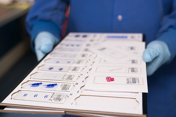
Section: A “section” refers to one full-face slice off of an FFPE block from a microtome. Standard practice is to place one section per slide unless otherwise requested in the Cutting Instructions of the ARUP requisition.

Serial section/ribbon: A string of sections is known as a serial section or tissue ribbon. This is how sections come off the microtome; they are then separated into individual sections and placed on a slide.

Frozen Specimens: Preparation and Embedding

Cryomolds are used to embed frozen specimens in optimal cutting temperature (OCT) compound. We use three different mold sizes to accommodate different tissue sizes: a standard size (25 mm x 20 mm x 5 mm), an intermediate size (15 mm x 15 mm x 5 mm), and a deep size (surface size is slightly larger than the standard, and depth is for particularly thick pieces of tissue).

Cryoprocessing refers to the process of preparing frozen tissue for embedding and cutting. As freezing tissue functions as a (physical) “fixative,” so to speak, the tissue itself does not need to be placed into a chemical fixative. Frozen tissue can be submitted either fresh, wrapped in saline-moistened gauze, on wet ice, or in a 30% sucrose holding solution.

OCT compound stands for “optimal cutting temperature” compound. This admixture of water-soluble glycols and resins is used to embed tissue specimens prior to frozen sectioning on a microtome-cryostat.

Frozen Specimens: Cutting/Sectioning

Frozen sectioning: Frozen specimens in OCT are cut on a cryomicrotome, in sizes ranging from 3 μm to 30 μm.

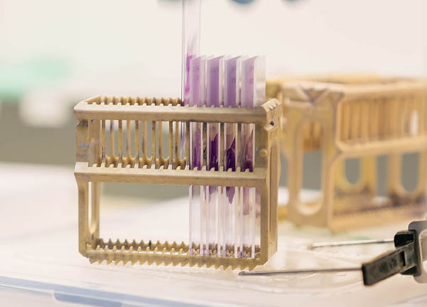
Slide: Slides are made of glass and serve as a mount for tissue sections. Sections stick to a slide when the slide is charged.

Coverslip is a plastic wrap that goes around a stained slide to protect it from environmental damage.
Administrative Terms

Project order refers to the totality of a submission. This phrase is more often used in accounting and billing contexts. The difference between the terms submission and project order is pragmatic. Whereas “submission” is used in reference to the physical handing in of materials, the phrase “project order” is used when referring to the nonphysical aspect of a submission, that is, it underscores the fact that the submission is an “order” made by the client. 

Requisition form is the official order form handed in along with a submission that includes the client number and project title, contact information, number of cassettes submitted, submission instructions, list of identifiers (block IDs), relevant order instructions, and additional comments.

Specimen(s) refers to the tissue itself. The term may also more broadly refer to the tissue and accompanying paraphernalia, such as the tissue along with the cassette, in Histowrap, etc.

Submission refers to all the components of a project order: the specimens, completed requisition form, list of specimens, and any other accompanying materials. The difference between the terms submission and project order is pragmatic. Whereas “submission” is used in reference to the physical handing in of materials, the phrase “project order” is used when referring to the nonphysical aspect of a submission, that is, it underscores the fact that the submission is an “order” made by the client.

Other Terms

Artefact: Carson describes an artefact as any “structure or substance not normally present but produced by some external force or action.” (Carson, 2009, p. 366) Examples include knife/cutting lines, misshapen cell morphology, and tissue floaters. Proper preparation and fixation are paramount for reducing associated artefacts, as these steps cannot be redone, whereas samples may be re-embedded if air bubbles are present or another section obtained if a preventable knife line is present.

Core punch: A core punch refers to punching out a cylinder of tissue from an FFPE tissue block and is also referred to as “punch” or “tissue punch.” It is different from “core,” which refers to a needle core biopsy. The standard size of a core punch is 2 mm, but we also offer 1-mm-sized punches upon request. If a different core punch size is needed (e.g., 3 mm, 5 mm, etc.), you will need to provide the punching tool. For research projects using archival human tissue, the approval of the medical director is required.

Needle core (biopsy) refers to cores of tissue taken with a needle during a biopsy.

Tube: A tube refers to the container that may hold a core punch or a scroll/shave.

Scroll/shave: Scroll and shave are interchangeable terms that refer to sections cut in such a way that the section curls up in the shape of a scroll. Scroll/shave is often used for DNA extraction.

Works Cited

Carson FL, Cappellano CH. Histotechnology: a self-instructional text. 3rd ed. American Society for Clinical Pathology Press; 2009.

Recommended Products

Tissue Wrap:

Histowrap by Obex, 2 x 3” Rectangle
Histowrap.com

Tissue Cassettes:

VWR Premium Tissue Cassettes (biopsy-routine size tissue)
https://us.vwr.com/store/product/4638105/vwr-premium-tissue-cassettes
VWR Mega Cassettes (30 x 40 x 11 mm)
https://us.vwr.com/store/product/7595491/vwr-mega-cassettes
VWR Mega+ Cassettes (whole mount 53 x 75 x 12 mm)
https://us.vwr.com/store/product/7595491/vwr-mega-cassettes
FAQs

Research Histology has provided answers to these frequently asked questions (FAQs). If you need more information, please contact the office at 801-213-4272, Monday–Friday, 8 a.m.–4 p.m.

FAQs Regarding Submission

How do I submit tissue?

Complete submission guidelines can be found in our Submission Guidelines section.

How do I submit fresh tissue for frozen projects?

Optimally, fresh tissue will be wrapped in saline-moistened gauze and placed in a conical tube labeled with an identifier. The conical tube will then be placed on wet ice for submission. Tissue should be stable for up to 6 hours.

Can I make a frozen submission if the tissue has been fixed in paraformaldehyde?

Yes, you can submit tissue to be frozen if it has been fixed in paraformaldehyde (but not if it has been fixed in formaldehyde). Prior to submission, the paraformaldehyde needs to be removed from the sample. Place specimen in 3% sucrose solution until tissue sinks, and then repeat in a 10%, 20%, and 30% sucrose solution. Lastly, place each specimen in a 30% sucrose solution in individual vials.

When can I drop off fresh tissue for frozen projects?

Please drop off fresh tissue for frozen projects no later than 3 p.m. Additionally, call the lab beforehand at 801-213-4272 to notify us that you will be dropping off fresh tissue.

Are there special submission guidelines for tissue types?

Any specimen containing bone needs to be decalcified (which ultimately improves the quality of the sections). Research Histology will provide this service for an additional charge. There is a box on your client requisition form to indicate that the tissue needs decalcification. When dropping off samples, please stamp your requisition form with the “DECAL” stamp that is adjacent to the drop-off box.

What do I do if my list of specimens does not fit on the requisition form?

If the number of specimens you are submitting exceeds the number of spaces on the requisition form, feel free to attach a list of specimens to the requisition form or use multiple requisition forms.

FAQs Regarding Services and Supplies Provided
       

Does Research Histology provide supplies, such as Cryomolds, cassettes, and Histowrap?

Research Histology does not provide any supplies to clients. We do recommend certain products, which can be found on our Recommended Products page.

Can I have my project rushed?

Research Histology does not rush projects.The lab operates on a first-come, first-served basis. The standard turnaround time (TAT) for routine projects is one business week (five business days). However, TATs can vary depending on current volumes. The current TAT is posted in the lab below the submission drop-off location. Large projects, projects involving special or immunohistochemical staining, and frozen projects will take longer than one week.

Does Research Histology gross my specimens?

Research Histology does not provide this service. All specimens need to be grossed and placed in a cassette prior to submission to Research Histology.

Does Research Histology cassette my specimens?

Research Histology does not provide this service. All specimens need to be in cassettes and labelled with a unique identifier before being submitted.

Does Research Histology transfer formalin-fixed tissue into the 70% ethanol solution?

Research Histology does not provide this service. Clients are required to submit all specimens in a 70% ethanol solution after fixing the tissue.

FAQs Regarding Turnaround Time (TAT)

What is Research Histology’s TAT?

Research Histology’s standard TAT is one business week (five business days). During particularly busy times, the TAT may be extended to a week and a half or two weeks. TAT is posted near our project drop-off box.

If my project requires only embedding, will the TAT be faster?

While it is possible that an embed-only project will be completed faster, it is not guaranteed. Factors affecting the TAT for an embed-only project include the number of embed-only submissions and staffing.

FAQs Regarding Technical Aspects of Projects

How thick are the scrolls cut?

Generally, scrolls are cut at 10 microns but Research Histology can cut any number of scrolls at the desired thickness as long as the instructions are indicated in the Cutting Instructions section on the requisition.

What size are punch kits?

Research Histology provides 1 mm and 2 mm punch kits for tissue punches. If a different size punch is needed, the punch kit needs to be provided with the submission. We require a circled hematoxylin and eosin (H&E) accompany the request indicating the area to be punched.

How do I indicate orientation for embedding/cutting?

The tissue orientation during embedding will determine the orientation of cutting. Orientation can be indicated in one of two ways: 1) by inking, or 2) by providing detailed instruction or images describing the required orientation. Place the side of the specimen you want cut first on the bottom of the cassette, which will be transferred during embedding directly to the bottom of the mold, which is the side that will be cut first .
casset

You can find more information on the Terms, Definitions, and Relevant Submission Guidelines sections.

How do I ink my tissue to indicate orientation?

Pat the desired area dry with gauze. Apply desired ink only to the area of interest and set using distilled vinegar. Return tissue to desired fixative.

FAQs Regarding Submission

How late is the lab open?

Research Histology is open from 8 a.m.–4 p.m., Monday–Friday. Projects can be placed in our drop-off box during business hours. If you have fresh tissue for a frozen project, it needs to be submitted no later than 3 p.m., as we freeze them immediately.