

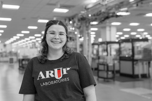

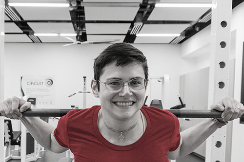
She didn’t feel math-y to start. Meghan Maughan, now an ARUP histotechnologist, liked science in high school and college, but felt her math skills held her back from science careers.
These days she works in a field that integrates biology, chemistry, histochemistry (identification and distribution of tissues’ chemical constituents), and immunology. On a daily basis, she prepares tissue for microscopic examination where a pathologist looks for tumors and discerns information about cellular changes and other alterations in cellular and organ development.
Meghan Maughan“I felt I was too old to pursue the long path of becoming a doctor. I told my husband I wished I was a scientist 200 years ago, when part of the job involved taking notes and drawing pictures. After I began to look at what regular histology labs do, creating special stains and dyes, sometimes gold and silver, and applying them to tissues—I liked that it involved a little inclination toward fine motor skills, the creative mind.”
Histotechnologist, Immunohistochemistry
Back in college, Maughan studied environmental studies and anthropology. Then she spent years as a grantwriter in homeless services and at the University of Utah’s Pioneer Theater Company.
How did she begin helping to diagnose cancer? Maughan talks about that with us and gives tips for others.
What is your current work?
In the Immunohistochemistry lab at ARUP, we work with antibodies—blood proteins that counteract a specific antigen.
Antigens are toxins or foreign substances that instigate the body’s immune response, particularly in producing antibodies. Inside bodies, antibodies combine chemically with “alien” substances including bacteria, viruses, and foreign substances in blood.
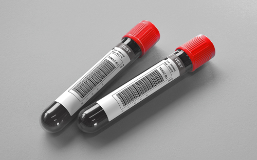
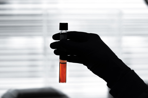
In our lab, we begin with patient tissue that has been fixed in formalin—a substance that fixes the tissue in place for examination, partly composed of formaldehyde in phosphate-buffered saline solution—and embedded in a block of paraffin wax. Using a microtome (a tool used to cut very thin slices of material for microscopy), we cut a section of the paraffin embedded tissue and mount it onto a slide. We then load that slide onto an automated stainer.
Once that’s on the stainer, we remove the paraffin wax from the patient tissue, and the methylene bridges formed during the fixation process are broken to expose epitopes (specific targets against which an individual antibody binds) and facilitate antigen-antibody binding. This last part is called retrieval. After the retrieval process, we apply a carefully prepared antibody dilution to the tissue, where it attaches to the antigen of interest. We then apply a secondary antibody to the tissue, linking the primary antibody to a reporter enzyme (a specific enzyme that researchers attach to a regulatory sequence in a gene of interest in bacteria or cell culture).
Then, we add chromogen to create a visual of the antibody/antigen complex, creating brown or red staining. Finally, we apply a counterstain to the tissue to aid in analysis. We remove the tissue from the automated stainer, run it through a series of graded alcohols to dehydrate it, then coverslip the slides and review them under a microscope, before sending them to the pathologists for analysis. We are constantly learning, thinking, and helping doctors diagnose cancer—all of that fascinates me.
What else is exciting?
If you’re a naturally curious, creative thinker, this can be a very good fit; there is always troubleshooting to do. When you’re diluting the antibodies, if you miscalculate the dilution ratio, your stains could fail. Or maybe you made your antibodies correctly, but the machine or another part of the process went afoul. Naturally, you have to work with standardized procedures, but if anything goes wrong, you have to think about where in the process that incorrect result could have been caused. Then you need to figure out how to fix it. We run a control with each tissue—so if the control fails, you’d look at various factors that could have caused that. That’s where being a creative thinker is helpful, and I find that appealing.
How did you change careers?
After about 10 years of grantwriting, I was getting burned out. It is very high stress, with low pay. I also wanted hands-on work, but writing about budgets and fundraising requests wasn’t hands-on. Because I was working at Pioneer and that was part of the university, I began taking classes. I loved an anatomy class, so I began to think about what I could do in anatomy. I felt I was too old to pursue the long path of becoming a doctor. But I told my husband I wished I was a scientist 200 years ago, when part of the job involved taking notes and drawing pictures. I began to look at what regular histology labs do, creating special stains and dyes, sometimes gold and silver, and applying them to tissues. I liked that it involved a little inclination toward fine motor skills, the creative mind. So I decided to take a few additional science classes (and some college algebra and statistics classes) to try to become a histotechnologist. I was fortunate, and my supervisor in Immunohistochemistry gave me an opportunity without prior experience in the field. She interviewed me and basically said, “Let’s take a chance.” So that worked out well for me.
Any advice you’d give a person considering this field?
If you’re curious and you’d like a hands-on job that impacts people’s lives but isn't working directly with patients and clinicians, histotechnology can be really rewarding. There are a lot of diverse opportunities within the field. In addition to working in a histology or immunohistochemistry lab like we have at ARUP you could work in a research lab or at a hospital. Or you could be a specialist who works for one of the companies that manufactures our machines and reagents—they are experts in the field and provide technical assistance with assay development and antibody optimization when needed. I love that there are so many opportunities in this field.
Catherine Arnold, Science Communications Writer
Related blogs
"Medical Lab Technician Devon Turner, Automated Core Laboratory (Q&A)"