


65-Plus Labs, But What Happens Inside?
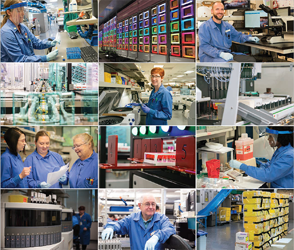
The more than 65 individual labs at ARUP Laboratories form a hive of variety. The labs are also highly centralized, resulting in quick turnaround times and efficient tracking, says Martha Bale, vice president, director of technical operations, ARUP. “Because everything is right here, we can send specimens between laboratories very quickly, not take a day to send them elsewhere. Tracking is faster, dramatically cutting the chance of misplaced specimens.”
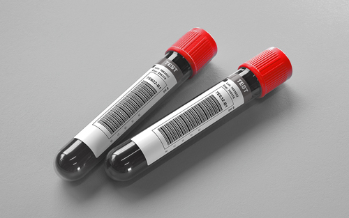
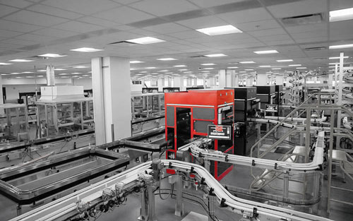
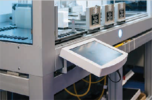
Many labs perform automated tests with amazing machinery, shuttling specimen tubes around tracks to ensure efficient processing. Is it expert engineering, Roald Dahl fiction, or both? (Fortunately, no one’s turned into a blueberry—yet.)
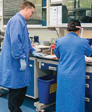
In some ARUP labs, there’s bustle and, more often than not, an automated result. In others, technologists work individually, carefully staining tissue or arranging paired chromosomes for examination, collaborating when needed to problem solve.
Despite these differences, lab technologists and managers have one thing in common: They notice. Technologists troubleshoot or look for patterns in the case of any unsuccessful result. Those who excel in their work have an eye for discerning irregularities and solving puzzles.
Want to take a look inside labs without donning white coats and protective gloves? Let’s get a glimpse into five of them here.
Automated Core/Automated Endocrinology Labs
State-of-the-art instruments are used to run about 180 categories of tests in these two labs. One of the primary tests in the Automated Core Lab is maternal serum testing, which looks for specific markers in amniotic fluid or serum/plasma from expectant mothers.
With tasks that vary from preparing test runs to analyzing data, a spectrum of specialists performs the work in both labs, says Ryan Greer, assistant vice president and group manager of Technical Operations, Chemistry.
The two labs’ high volume of tests makes them ideal places for technologists or technicians, some of whom have high school diplomas and are working on their medical laboratory science (MLS) degrees, to gain broad experience in traditional chemistry and laboratory work. Others have a place there, too. Technologists with MLS degrees can perform tests, and scientists with advanced degrees perform data analysis, says Greer.
Automation’s objective, he says, is to take testing that traditionally relied on a medical scientist to interpret and make judgments, and standardize the analytic process to achieve consistency. Test result accuracy is typically higher with automation, and smaller staffs can run large volumes on a 24/7 schedule, says Greer.
Brainpower still has a place in automated labs, though. “I’d like to debunk a myth that in automated labs, you just put samples in, push ‘go,’ and walk away,” notes Greer. Being able to load the analyzer and walk away frees staffers for other tasks, such as scrutinizing the data, he says.
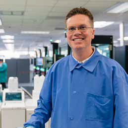
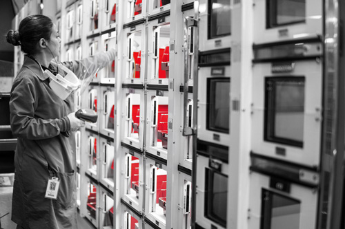
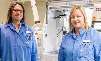
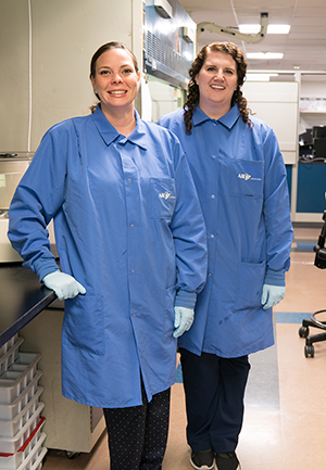
"A spectrum of specialists performs tasks that vary from preparing test runs to analyzing data in the Automated Core and Automated Endocrinology labs."
Ryan Greer, Assistant VP and Group Manager, Technical Operations, Chemistry
Technicians and technologists who excel “have an eye for detail, the ability to think critically, and can find subtle patterns and nuances in huge amounts of data—all of which makes them very successful at catching and understanding problems in data,” says Greer.
Cytogeneticists have a big job looking at small details.
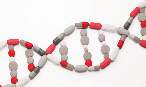
At ARUP, the Cytogenetics Lab performs two types of analysis: chromosome and fluorescence in situ hybridization (FISH). Using FISH, technologists prepare short sequences of single-stranded DNA called probes to match an area of the gene for which the researcher is looking. Fluorescent dye colors are attached to the probes to differentiate them. Researchers then compare the probes with the dual-stranded actual DNA to find differences. They can perform comparisons on nondividing cells that are between phases.
A chromosome analysis involves meticulously scrutinizing chromosomes from patient samples for gains, losses, or rearrangements of genetic material.
Through training and experience, technologists performing these tests develop a comprehensive understanding of each chromosome’s structure and its associated banding pattern. Some abnormalities are easily recognizable, such as a gain of an entire chromosome 21, which results in Down syndrome. Other abnormalities may require a high level of skill and experience to detect. This is especially true for oncology cases, in which only a couple of the 20 cells analyzed per case may be abnormal.
Identifying a low-level, abnormal clone takes serious, close examination. If found, such a detail determines the specific type of cancer present and predicts disease course. It can even help to establish a course of treatment and aid in the monitoring of treatment effectiveness. However, the necessary analytical ability isn’t developed overnight. “It typically takes three to five years of repetitive chromosome analysis training before a technologist is truly adept at analysis,” notes Brandon Chandler, technical supervisor, New York, in Cytogenetics at ARUP.
A few things happen before all that analysis, though. First, samples go through extensive processing in the wet lab. A technologist puts specimen cells into a wet media in which the cells can actively divide. The ideal result is many dividing (mitotic) cells. After a designated incubation period, cultures are harvested to produce a fixed cell pellet. Transferring the metaphase cells (at a stage of mitosis in which chromosomes are condensed and microscopically visible) from the pellet to slides takes significant skill, says Chandler.
The goal is to get the chromosomes in each metaphase cell to spread as much as possible without rupturing the cell membrane. It’s necessary to have a steady hand in dispensing the sample onto the slide, and temperature and humidity must be tightly controlled. The slides are stained, then scanned with a digital microscope system.
At that point—ta-da!—it’s time for analysis. Special software similar to that used for photo editing helps align chromosome pairs. Then they are ready to be evaluated carefully for aberrations. A straightforward case can be analyzed by an experienced technologist in less than an hour; however, a complex, abnormal case can keep an analyzer busy for several hours. Chromosome analysis is both challenging and fun, asserts Chandler, because you never know which type of abnormality you will encounter. Each cell is a unique puzzle waiting to be solved.
All that careful scrutiny can have a very cool conclusion, says Chandler. “If you do find that needle in the haystack in an oncology case, you’re helping to identify the specific abnormality that a patient has, and you’re helping a physician to zero in on the proper treatment. It’s rewarding to know that your efforts have a direct impact on someone’s life.”
Microbial Immunology Lab
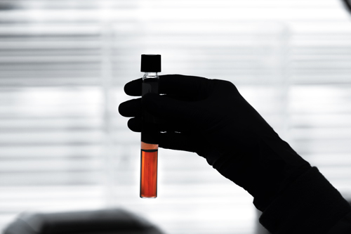
With a focus on infectious diseases, Microbial Immunology (Micro Imm) itechnologists sometimes do work that makes the news.
For example, this lab tested the blood of 457 Olympic athletes returning from the 2016 Summer Games in Rio de Janeiro for a University of Utah study reported last fall. The study’s surprising results showed that none of the tested Olympians had contracted the Zika virus.
One of the lab’s main tests is for Lyme disease; it also performs herpes tests and—less in the news, but in high demand for screening healthcare staff—tuberculosis tests. All in all, Micro Imm tests for 19 conditions, performing about 70,000 tests a month.
On a typical day, technologists arrive in the lab and check the schedule to learn which tests they’ll be running. Some tests begin with enzyme-linked immunosorbent assay (ELISA) methodology, using a microtiter plate of multiple indentations, or wells. Into each well, an antigen (a toxin or other foreign substance that causes an immune response in the body, especially antibody production) has been placed. The technologist adds the patient specimen to the same well.
In a test for Lyme disease, if Lyme antibodies are present in the specimen in the well, the antibody attaches to the antigen. The technologist rinses the serum and any antibody stays attached. Adding a chemical at this point produces a color reaction; the more antibody present in the specimen, the more intense the color reaction will be.
If the ELISA test is positive and a reflex test has been ordered, the technologist runs an immunoblot test, in which strips of paper with antigens attached are used to confirm whether antibodies are present for bacterial antigens in question.
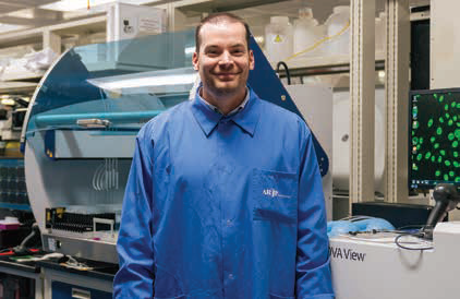
After working as a veterinary technician and loving the job’s lab work, Kathryn Olson, technical supervisor, talked with a microbiologist who advised her to get into medical technology. “That was the first I’d heard of it.”
Working in Micro Imm equals happiness for Kathryn Olson, technical supervisor in that lab. She chose lab work after finishing a bachelor’s degree in medical laboratory science and decided to obtain a master of science degree as well “because I loved the work so much.”
Parasitology and Fecal Testing (PAFT) Lab
In PAFT, the goal is—you guessed it—to find out if a clinging monster (that is, a parasite) lurks within. If one is found and identified, the illness it causes can be treated more effectively. Parasites can potentially infect almost any part of the human body, inhabiting fluids, tissue, etc. “That makes it always different for our lab. We get specimens from the eye, the blood; things that attach to skin, like ticks, or from the intestinal system, like tapeworms,” says Kristin Case, technical supervisor, PAFT.
The lab tests a range of matter: stool, skin scrapings, eye swabs. “We’re not just blood work in Parasitology,” Case notes. Hundreds of specimens are tested in batches each day.
Technologists rotate through several benches in PAFT every day. They analyze parasites using microscopes, and use testing kits and advanced instruments to analyze fungal elements and viruses. If someone is having trouble identifying an organism, the team works together to identify it. “Some are more experienced at identifying parasites and organisms than others, so we learn from each other every day,” says Case.
Tests performed in Parasitology include the following:
- Fungal antigen testing. This uses a methodology called enzyme-linked immunosorbent assay (ELISA). Technologists receive kits from manufacturers, then pipette a small amount of patient specimen into a small tube or well and add certain reagents. There may be a reaction if the antigen from the tested fungus is present.
- Fecal chemistry. Several tests are performed to analyze stool. For example, a technologist can look for fat in stool. If present, fat can be a sign that a person’s gastrointestinal tract is not absorbing fats or other nutrients properly.
- Ova and parasite examination. Technologists read stained slides using a microscope, looking for eggs, cysts, or other signs of parasites.
PAFT is absorbing to technologists day after day, says Madison Sant, senior medical technologist in that lab. “It’s an intriguing medical field, analyzing a patient’s specimen. You’re figuring out the mystery of what’s wrong with the patient. Even though we’re not the doctor or nurse in the room with the patient, we’re able to help the patient by identifying what’s wrong.”
Her parasitology training and ARUP’s connection with the University of Utah meant Kristin Case, technical supervisor, was able to train medical workers in China and Africa to be better parasitologists—while she helped collect data for the U of U.
University Hospital Lab
For the University of Utah Hospital, the Mountain West’s only academic medical center, this laboratory processes “nearly anything and everything that a hospital needs,” says Daniel Savage, technical supervisor of University Specimen Processing at ARUP, noting that most physicians’ diagnoses result from lab work. He isn’t joking; about 55,000 tests are performed monthly in the lab.
Samples come in with a need for speed from the Emergency Department, Intensive Care Unit, and other high-intensity areas. A test called Complete Blood Count with Platelet Count and Automated Differential (CBCAD) looks for hemoglobin, hematocrit, platelets, and white cell count differential. It’s “a quick look inside a patient’s blood. It quickly lets a physician know how to treat the patient,” says Savage.
Other specialty areas include:
- Therapeutic drug tests, which determine whether a patient taking a medication may need an increase or decrease in dosage
- Coagulation (coag) testing, which monitors therapy with anticlotting drugs like heparin so that doses can be adjusted accordingly
- Testing of body fluids, such as cerebrospinal fluid (CSF) found in the brain or spinal cord, or pleural (lung) fluid cell counts, to determine white blood cell abnormalities for Huntsman Cancer Institute and University Hospital
- Flow cytometry tests for specific biomarkers and proteins on abnormal cells. This helps identify which complication or disorder the patient is dealing with and the direction for treatment.
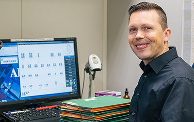
"Everything in the hospital gets funneled to us—the lab. Doctors can’t treat or do anything until they get results back and learn what to do next.”
Daniel Savage, Technical Supervisor, University Specimen Processing
The busy, intense world of a hospital lab is an exciting place to make a difference, says Savage. “The test numbers show how important lab testing is. It is how we can look inside a person’s body and figure out what’s going on, so it is absolutely vital.”
 HOME
HOME